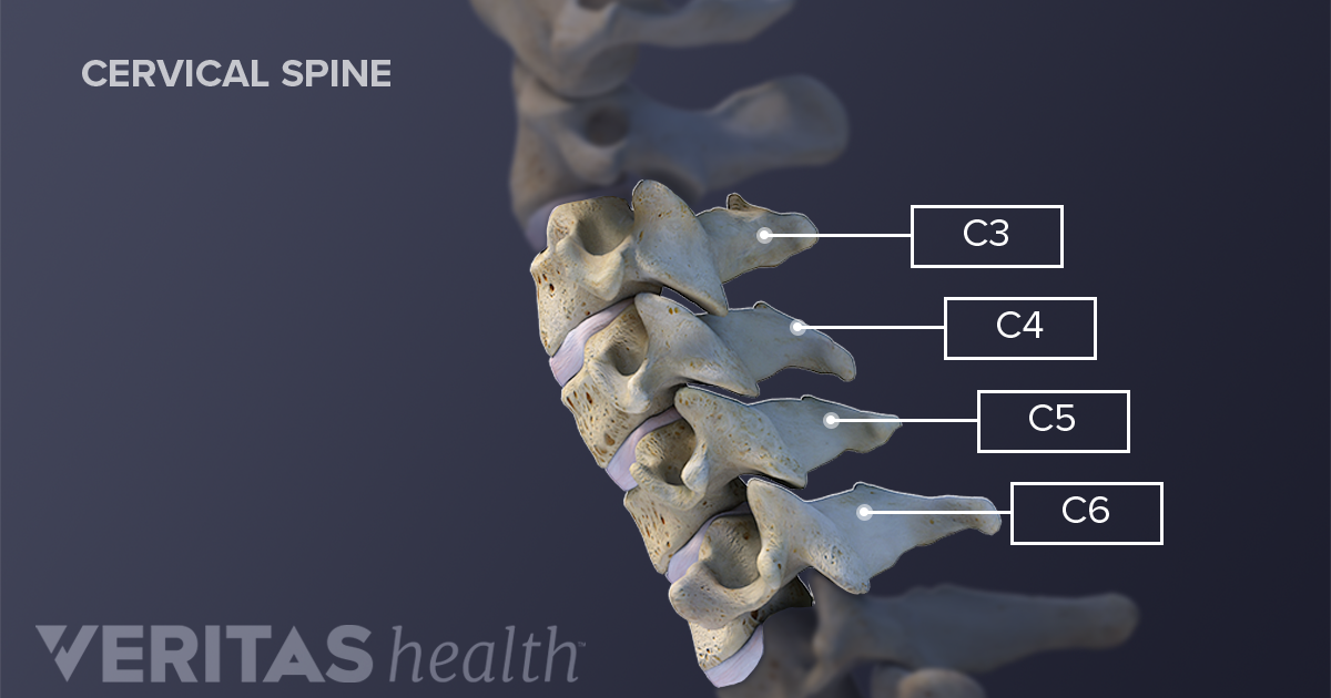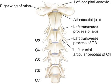
Its spinous process, on the other hand, is smaller than in the other lumbar vertebrae with a wide, four-sided shape that comes to a rough edge and a thick notch. The fifth lumbar vertebra is distinct from the L1-4 vertebrae in being much larger on its front side than in the back. Unlike the cervical vertebrae in the neck, the lumbar vertebrae lack the transverse foramina in the transverse processes, and also lack facets to either side of the body. The transverse processes form important connection points for many muscles, including the rotatores and multifidus muscles that extend and rotate the trunk. Natural fusions may occur in the neck and are often found in combination with disc degeneration, severe osteoarthritis and. On the left and right lateral sides of each vertebra are the short, triangular transverse processes. Naturally fused vertebrae can occur virtually anywhere in the spine, with the exception of the sacrum and the coccyx, which already demonstrate organic fusion of the vertebral bones by natural design in a typical anatomy. It serves as a connection point for the muscles of the back and pelvis, such as the psoas major and interspinales. The spinous process extends from the posterior end of the arch as a thin rectangle of bone. The vertebral foramen is a large, triangular opening in the center of the vertebra that provides space for the spinal cord, cauda equina, and meninges as they pass through the lower back.Įxtending from the vertebral arch are several bony processes that are involved in muscle attachment and movement of the lower back. The arch surrounds the hollow vertebral foramen and connects the body to the bony processes on the posterior of the vertebra. Posteriorly the body is connected to a thin ring of bone known as the arch. A cylinder of bone known as the vertebral body makes up the majority of the lumbar vertebrae’s mass and bears most of the body’s weight. The lumbar vertebrae are the some of the largest and heaviest vertebrae in the spine, second in size only to the sacrum. The jelly-like nucleus pulposus acts as a shock absorber to resist the strain and pressure exerted on the lower back. The outer layer of the intervertebral disk, the annulus fibrosus, holds the vertebrae together and provides strength and flexibility to the back during movement. Together they create the concave lumbar curvature in the lower back.Ĭonnecting each vertebra to its neighboring vertebra is an intervertebral disk made of tough fibrocartilage with a jelly-like center. Each cell type was generated from four different healthy control lines.The lumbar vertebrae are stacked to form a continuous column in order from superior (L1 or first lumbar vertebra) to inferior (L5 or fifth lumbar vertebra).

The samples included 3 different cell types: iPSC macrophage precursors, iPSC microglia grown in monoculture and iPSC microglia grown in co-culture with iPSC motor neurons and extracted using CD11b MACS. We sequenced a total of 12 samples, with 2 sequence runs per sample. This novel and authentic human model system facilitates the study of physiological motor neuron-microglia crosstalk and permits the investigation of non-cell-autonomous phenotypes in amyotrophic lateral sclerosis. Further, they express key amyotrophic lateral sclerosis-associated genes and release disease-relevant biomarkers. Sometimes, the inflammation may also affect the sites where ligaments and tendons attach to the bones of the spine. It may be related to wear and tear, autoimmune disorders, infection and other conditions.

Co-cultured microglia express key microglial markers and transcriptomically resemble primary human microglia, have highly dynamic ramifications, are phagocytic, release various cytokines and respond to stimulation. Spinal arthritis is inflammation of the facet joints in the spine or sacroiliac joints between the spine and the pelvis. Here, we combine both cell lineages and establish a novel co-culture of iPSC-derived motor neurons and microglia, which is compatible with motor neuron identity and function. In our previous work, we have established protocols for the differentiation of iPSC-derived spinal motor neurons and microglia. GEO help: Mouse over screen elements for information.Ĭo-culture of iPSC-derived microglia and spinal motor neurons as a novel model for amyotrophic lateral sclerosisĮxpression profiling by high throughput sequencingĪmyotrophic lateral sclerosis is a fatal neurodegenerative disorder primarily characterized by motor neuron degeneration with additional involvement of non-neuronal cells, in particular, microglia.


 0 kommentar(er)
